Selected work · 05
Graphene Transfer
Preserve a graphene film through wet transfer, then ask what the measurements actually show.
Explore the engineering ↓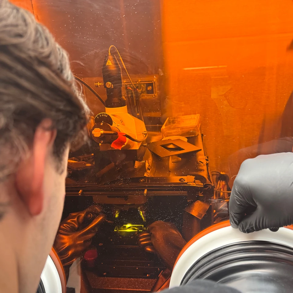
- My role
- Joint experimental work and presentation with Hunter
- Status
- Transferred samples characterized
- Focus
- Wet transfer · Raman · AFM
Result & scope
The presentation reports G (~1580 cm⁻¹) and 2D (~2700 cm⁻¹) Raman features in transferred material. Layer count and defect-free transfer are not established.
- 01
Support
Use PMMA to support CVD graphene on copper.
- 02
Transfer
Etch the copper and move the supported film to a new substrate.
- 03
Locate
Inspect optical contrast and choose regions of interest.
- 04
Characterize
Compare Raman signatures with AFM topography on exfoliated flakes.
Evidence from the project
Inside the work.
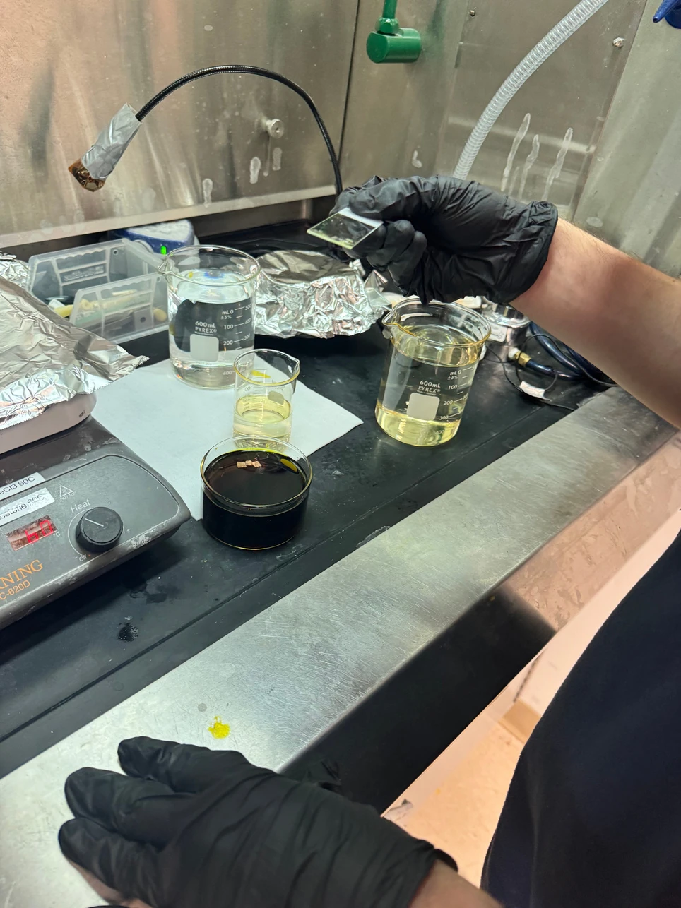
Project photograph from the transfer presentation; handling and sample preparation are part of the experiment.
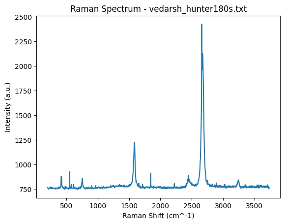
The supplied plot shows the reported G and 2D features. No new fit or raw-data reconstruction is added.
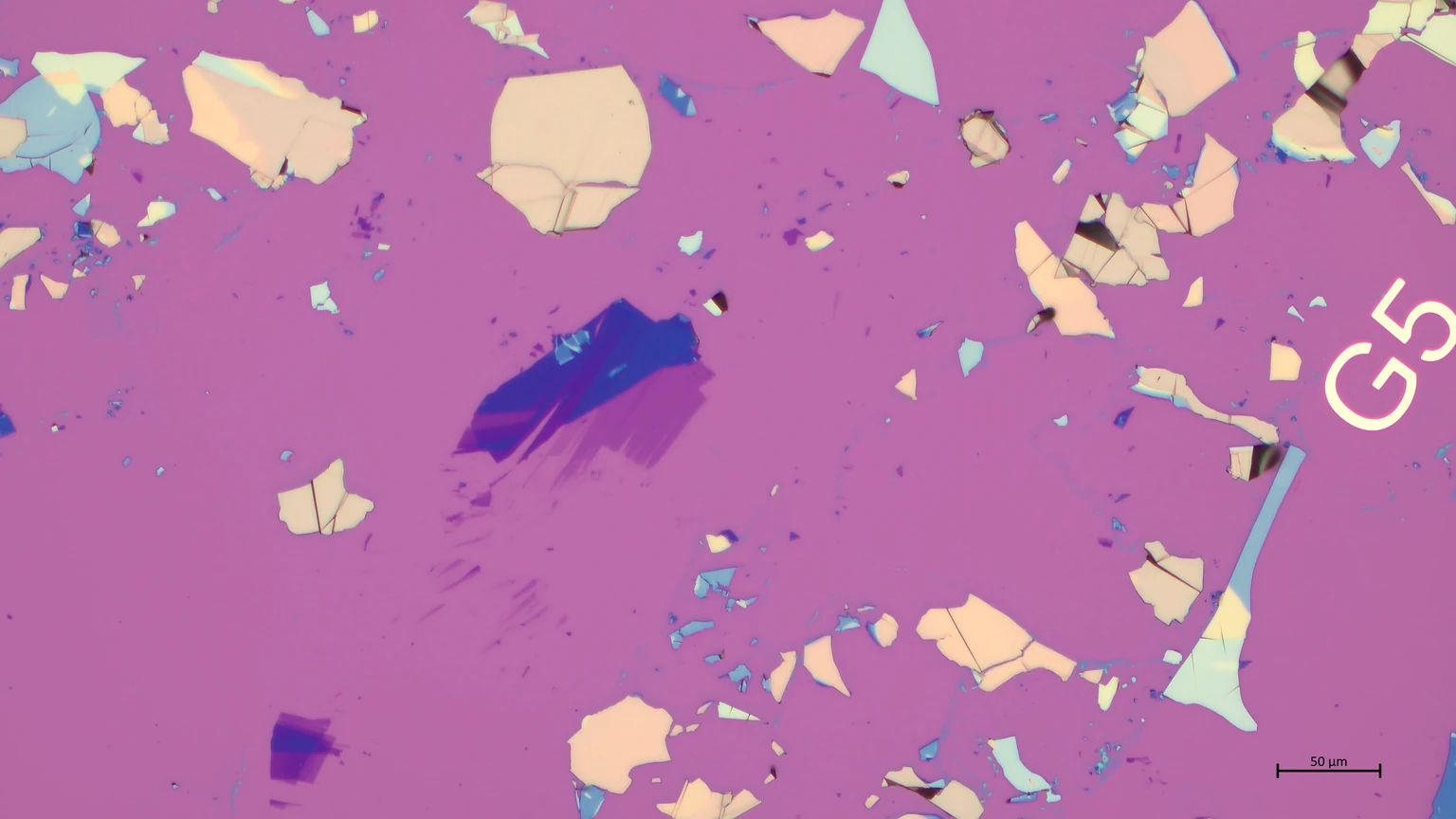
Variation in size and contrast motivates region-by-region characterization.
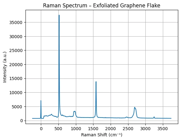
D, G, and 2D features provide a different structural view from the transferred-film sample.
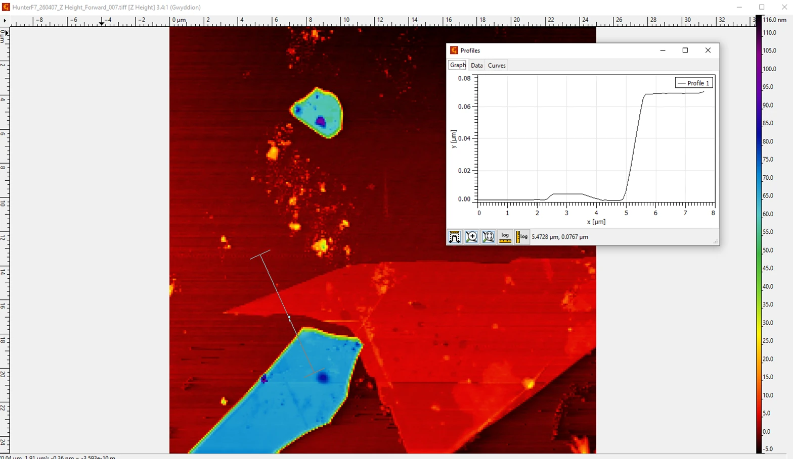
Topography and the step trace support a distinct layered structure; they do not prove uniform monolayer material.
Two sample routes, different questions
The joint presentation separates a CVD wet-transfer route from mechanical exfoliation. Wet transfer aims to preserve a continuous film; exfoliation produces isolated flakes with variable size and thickness. Those routes should not be treated as equivalent samples.
Transfer and structural evidence
The CVD process uses a PMMA support layer while copper is removed, followed by transfer to a new substrate. Raman spectra are used to check for graphene-like structural signatures after processing. The source reports artifact removal before interpretation; the page retains the supplied plot rather than reconstructing unprovided raw data.
Raman and AFM answer complementary questions
The transferred-film spectrum contains G and 2D features. The exfoliated-flake spectrum also includes a D feature near 1350 cm⁻¹, consistent with defects. Optical images guide selection, while AFM reveals topography and a step edge.
Limits of the conclusion
These observations support graphene identification and a distinct layered structure. They do not establish uniform monolayer coverage, a numerical defect density, or a transfer yield. The presentation describes nonuniform flakes and potentially multiple layers; those limitations remain part of the result.
Read the source
Technical records.
About the evidence
Graphene Transfers, joint presentation by Veda and Hunter: wet transfer, Raman, optical microscopy, and AFM sections. Original plots are retained.
Continue exploring.
← Back to Selected Builds